Beam-Foil Spectroscopy System
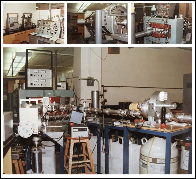
Designed and constructed a beam-foil spectroscopy system at the UTA accelerator facility to study the structure of O, N, and S ions. A gas-handling system was built to allow injection of H2, He, O2, N2, Ne, and SF6. An energy analysis op-amp circuit was designed to adjust the Van de Graaff potential to maintain precise velocity of the ions. A high-vacuum target chamber was built which incorporated stepper motors to select and move the target foils. An Acton VM-502 monochromator and Hamamatsu R595 electron multiplier were used to measure the radiation emitted from the foil. The foil was translated up to 10 cm with respect to the slit of the monochromator in order to measure the change in intensity of the fluorescence from the ion beam as a function of distance from the foil. The data from these intensity measurements determined the oscillator strength, which is the probability of emission of radiation for transitions between energy levels of the atom. A microcomputer system interface was designed which provided instrument control, data acquisition, and analysis.
Vacuum-Ultraviolet Spectroscopy System
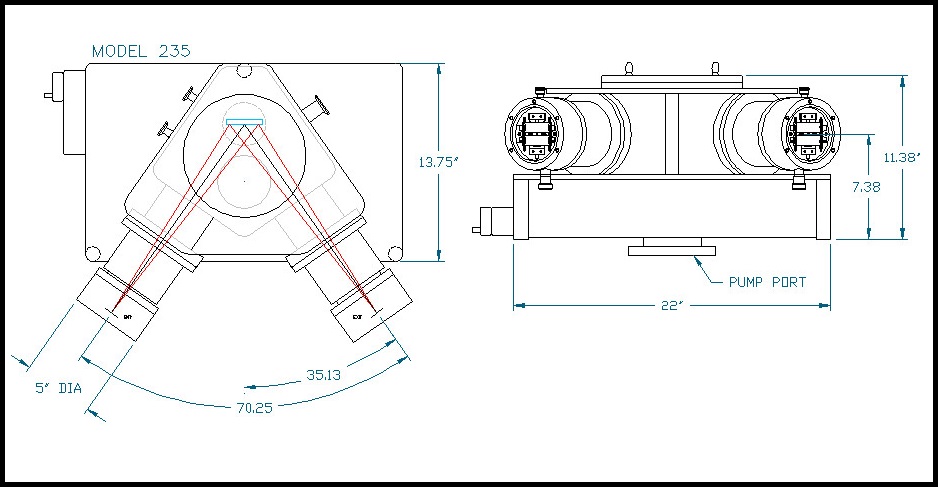
Designed and assembled a vacuum-ultraviolet (VUV) spectroscopy system (10 nm – 200 nm) using a McPherson 235 Seya-Namioka Monochromator. The system used a diffusion vacuum pump and pump controller system to achieve pressure of 1×10^-6 Torr. Various low-pressure VUV light sources were designed and built, which included a hollow cathode lamp, 27 MHz RF discharge lamp, and electron-gun using a modified 6BN6 vacuum-tube. Output photons from the monochromator were detected using a Spiraltron electron multiplier SEM 4219. A low-noise photon-counting circuit and computer interface were built for detection of low intensity VUV radiation.
Ultraviolet Spectroscopy System for Biology Research
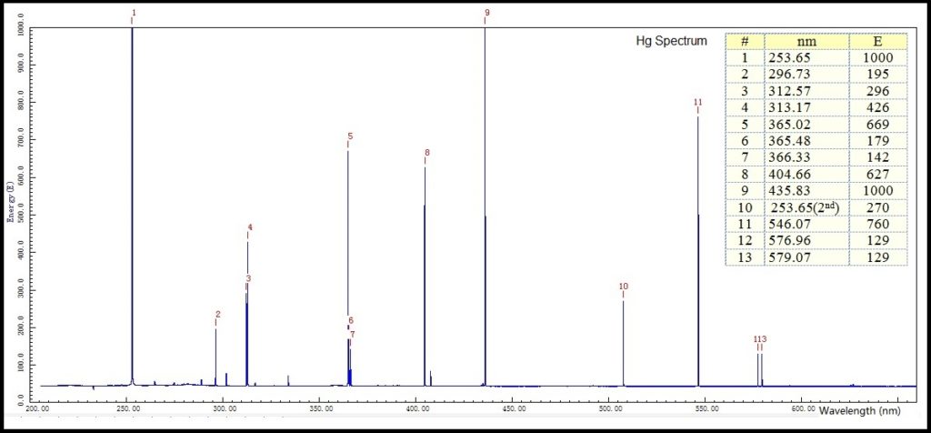
Implemented a UV spectroscopy system for biological applications to study how photoreaction enzymes repair damaged DNA by inducing ultraviolet light. Photoreactivation is the reversal of the harmful effects (growth delay, mutation, cell death, cancer) due to far-UV radiation (200-300 nm) on organisms by exposure of the organism to near-UV/blue light (300-500 nm). The photoreaction enzyme was discovered by Dr. Claud S. Rupert while he was at Johns Hopkins University. He continued this research at UTD and was on my doctoral committee. The system that I developed utilized a Lambda Hg lamp, Jarrell-Ash Spectrometer and a Princeton Applied Research optical multichannel analyzer OMA-1205 for spectral analysis.
Combustion Monitoring
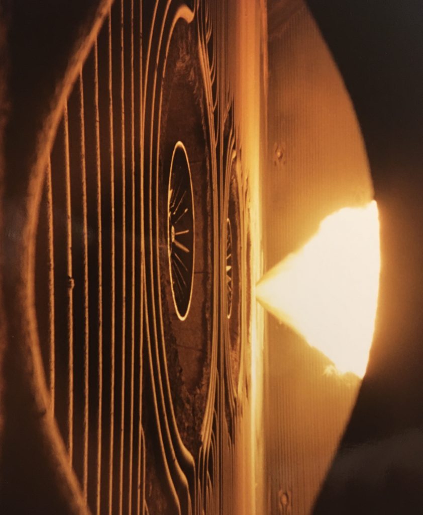
Designed various combustion monitoring systems which were implemented in the oil, gas, and coal power plant industries. These systems consisted of spectroscopic instrumentation and fast-response optical detection systems that monitored the infrared, visible, and ultraviolet flame emissions from the combustion processes. Measurements were obtained with Oriel 77250 Series 1/8 m monochromators, Newport and Melles Griot UV, VIS, and NIR bandpass filters, and Si, Ge, InGaAs, and PbS sensors. Flicker frequencies were extracted from the spectral data by performing real-time Fourier transforms on the voltage-time data. This provided information on the air-fuel ratios and combustion efficiency, and provided discrimination studies of individual flames in multi-burner systems.
Flame Detector for Gas Turbine Systems
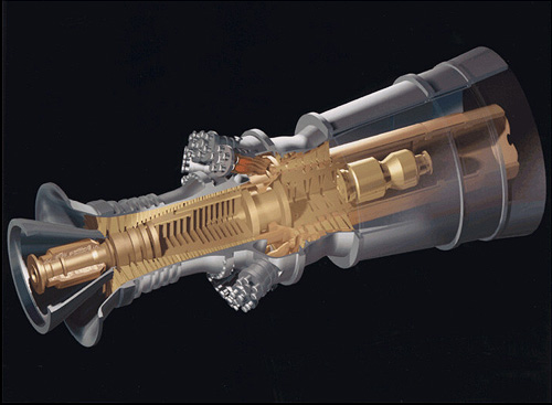
Developed a flame detection system for gas turbine systems which utilized an extended-range PbS sensor (New England Photoconductor). A high-temperature Fiberguide fiber-optic bundle and focusing lens assembly was integrated into the gas turbine combustion section. The remotely mounted optical sensor included a high-gain op-amp circuit and transmitted the signal to an analog high-speed signal processor. The high-speed signal processor had a selectable bandpass filter to respond at the peak flicker frequency which allowed precise response to the internal combustion. This provided accurate signal discrimination between combustion and background heat radiation allowing fast cutoff of fuel supply on flameout.
Helium Cell Absorption
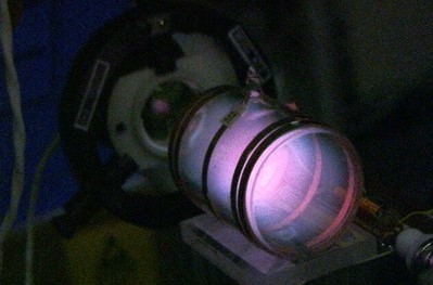
Designed the optical characterization system for helium cells used in high sensitivity laser magnetometer systems for NASA, JPL, NSF, and DOD. The helium cell sensor can be considered to be a variable density optical filter. The sensor utilizes field-dependent light absorption to sense the magnetic field. In the test-setup, helium in the absorption cell (1 Torr) was excited by an RF discharge (27 MHz) to maintain a population of metastable atoms. Circular polarized laser radiation (1083 nm) passed through the cell and produced an orientation of the metastable helium atoms with a net magnetic moment. The emergent radiation was detected by an IR detector (InGaAs) and the preamplifier signal output was a measure of the absorption in the cell. A Lorentzian function was used to model the spectroscopic absorption data with amplitude and width coefficients characterizing the cell.